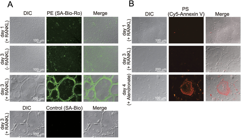
Figure 2. Increase of PE on the cell surface during osteoclast differentiation.
Osteoclast precursors were cultured for 1 day or 3 days with M-CSF in the presence or absence of RANKL, and then exposure of PE (A) and PS (B) on the cell surface was assessed. (A) PE staining with SA-Bio-Ro. The cells were treated with cell-impermeable SA-Bio-Ro for 30 min and fixed. SA-Bio-Ro bound to the cell surface was stained with a FITC-conjugated anti-streptavidin antibody. DIC, difference interference contrast. For control, differentiated osteoclasts were treated with SA-Bio and immunostained. (B) PS staining with Cy5-conjugated annexin V. For positive control, differentiated osteoclasts (day 3) were cultured for an additional 1 day with 10 μM alendronate to induce apoptosis.

