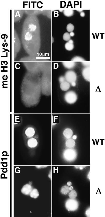
Figure 7.
Expression of methylated H3 Lys 9 and Pdd1p. (A–D) Localization of methylated H3 Lys 9. Wild-type (A,B) or DCL1 knockout RI strain (C,D) at nuclear alignment stage (12 h post-mixing) were stained with anti-methylated H3 Lys 9 antibody (A,C) or with DAPI (B,D). (E–H) Localization of Pdd1p. Wild-type (E,F) or DCL1 knockout RI strain (G,H) at nuclear alignment stage were stained with anti-Pdd1p antibody (E,G) or with DAPI (F,H). All photos share the scale bar shown in A.

