
Figure 6.
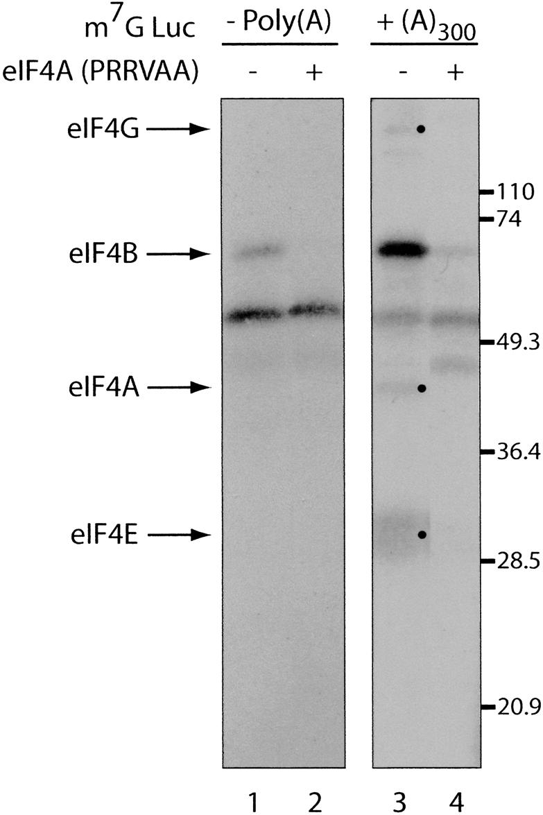
The poly(A) tail stimulates initiation factor binding to the m7G cap. Cap-labeled Luc(A)– (left panel) and Luc(A)+ (right panel) mRNA were incubated with rabbit reticulocyte lysate in the absence (lanes 1,3) or presence (lanes 2,4) of an eIF4A dominant-negative mutant (PRRVAA, 100 μg/mL) (Svitkin et al. 2001b), and subjected to UV cross-linking. Samples were analyzed by SDS-15% PAGE and bands were revealed by autoradiography. Bands corresponding to initiation factors are indicated with arrows on the left and bullets. The positions of molecular weight markers are indicated on the right.