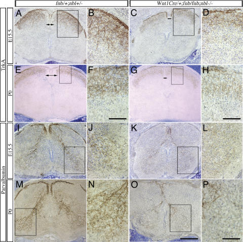
Figure 3.
Defects in nociceptive and proprioceptive innervation in the spinal cord of Wnt1Cre/+;fnb/fnb;nbl–/– mice. Nociceptive sensory afferent fibers, highlighted by the TrkA antibody, are examined at E15.5 and P0. (A–D) At E15.5, exuberant ingrowth of TrkA+ fibers is identified in the developing dorsal horns of control spinal cord (A,B). Some of the TrkA+ fibers extend into the intermediate layers of the dorsal horn. In contrast, the intensity of TrkA+ fibers is much reduced in the spinal cord of Wnt1Cre/+;fnb/fnb;nbl–/– mice. Note the reduction in the size of dorsal funiculus (DF, double arrows) in mutant spinal cords. Comparisons are made at the similar of levels of the spinal cord. (E–H) By P0, TrkA+ fibers form a compact layer of innervation at the dorsal horn of control mice, whereas the staining intensity of TrkA+ fibers continues to be reduced in the mutants. Compare the staining intensity in panels F and H. Proprioceptive fibers are labeled using the antibody that recognizes parvalbumin. (I–L) As early as E15.5, parvalbumin+ fibers form compact bundles in the dorsal roots (arrows) and dorsal funiculus (arrowheads) and project into the ventral horn of the spinal cord (I,J). In contrast, only sparse number of parvalbumin+ fibers are identified in the dorsal funiculus of the mutant spinal cord (K,L). The control spinal cord at P0 shows intense parvalbumin+ fibers in the Ia afferent projection to the ventral spinal cord. In mutant spinal cord; however, the number of parvalbumin+ fibers remains significantly reduced and only spare parvalbumin+ fibers are identified in the ventral horn. Bars: F,H, 25 μm; O, 200 μm; P, 50 μm.

