Figure 5.
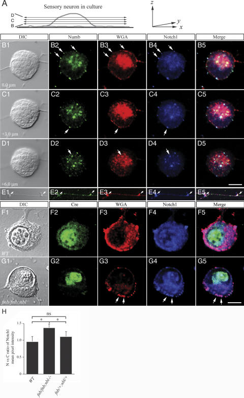
Loss of Numb and Nbl leads to a reduction in endocytic vesicles. (A) Schematic diagram depicting the confocal planes obtained from a sensory neuron in culture, which are presented in B–D. (B–E) Colocalization of Numb endosomal marker WGA and Notch1 ICD in the cytoplasm and axons of sensory neurons. Numb was present in endocytic vesicles and colocalized with Notch1 ICD in the wild-type sensory neurons (arrows in panels B–D). Interestingly, the colocalization of Numb and Notch1 ICD was also present in varicosities along sensory axons (E). (F,G) Deletion of numb, using HSV-iCre, in the nbl null background resulted in a marked reduction of WGA-positive endocytic vesicles in the cytoplasm (G3), without affecting the presence of endocytic vesicles in the cell surface. The presence of intracytoplasmic Notch1 vesicles was reduced (G4). Most of the Notch1 protein was present on the cell surface (arrows in G4). DIC images in B1–G1 were presented to define the boundary of the nuclear membrane (Q,V). (H) Quantitative analyses of Notch immunofluorescent intensity in nucleus and cytoplasm. Ratio of nucleus-to-cytoplasm Notch was determined using Leica Confocal Software (for details, see Materials and Methods). Whereas expression of Cre recombinase did not alter the nucleus-to-cytoplasm ratio of Notch in either wild-type or fnb/+;nbl/+ neurons, it resulted in a modest increase of such ratio in fnb/fnb;nbl/nbl neurons. Student's t-test, *p < 0.05; ns indicates not significant. Bars: D5, 7 μm; G5, 5 μm.

