Fig. 2.
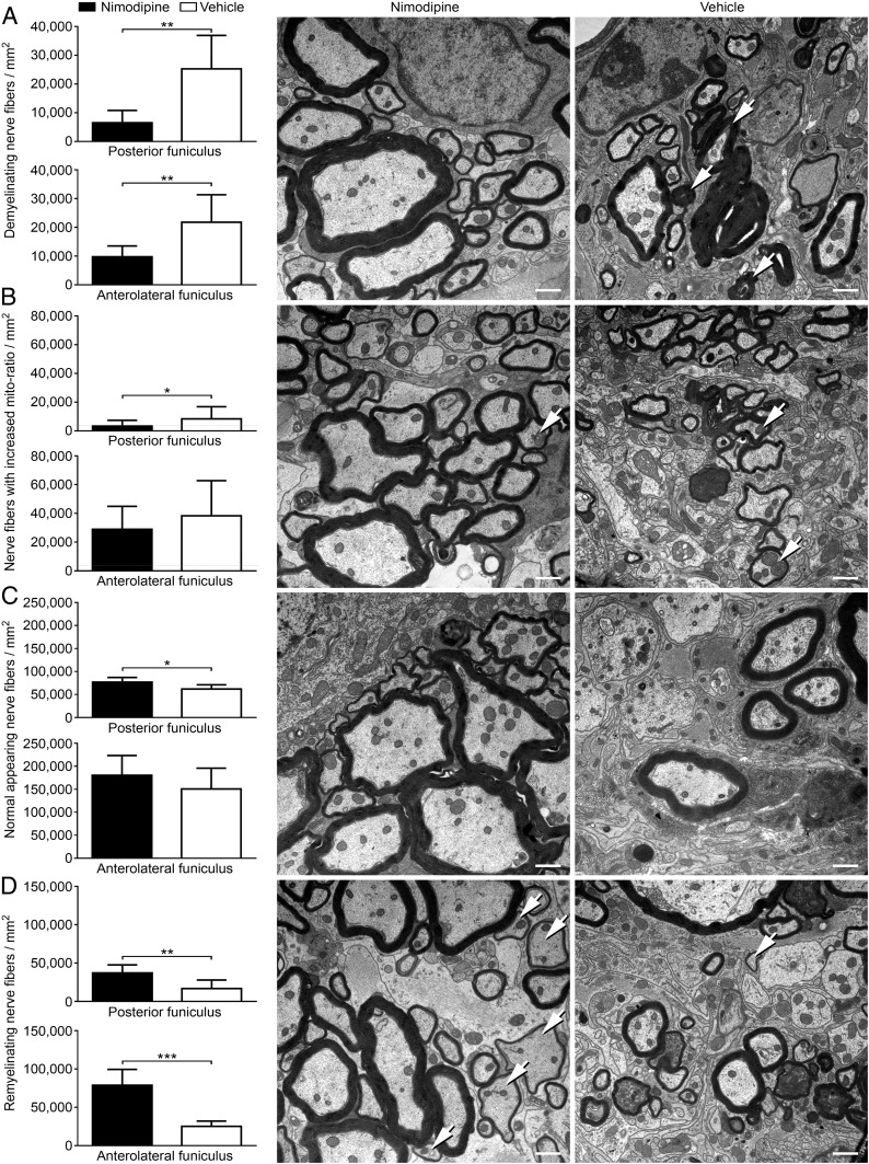
Decreased nerve fiber pathology and increased remyelination in nimodipine-treated mice. The posterior and anterolateral funiculus of nimodipine-treated (n = 12) and vehicle-treated (n = 9) mice were analyzed ultrastructurally. For these analyses mice were killed on day 68 or 72, respectively, after immunization. The following parameters were analyzed per square millimeter: demyelinating nerve fibers (A), the number of nerve fibers with increased mito-ratio (B), axonal loss (C), and the number of remyelinating nerve fibers (D). (Scale bars: 1 µm.) Arrows indicate representative features of the respective panels. *P ≤ 0.05; **P ≤ 0.01; ***P ≤ 0.001 by two-tailed, unpaired Student’s t test.

