Fig. S5.
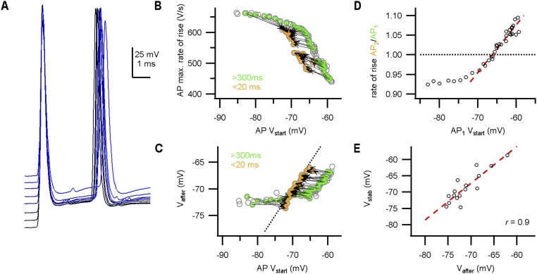
The AP stability potential Vstab correlated with Vafter in slice recordings. (A) During constant-current injections, the calyceal axon was stimulated at the midline to elicit a doublet with 3-ms interval. APs from a P5 calyx are aligned on the peak of the first AP. Stimulus artifacts are subtracted. (B) AP maximal rate of rise against the onset potential. Doublets are indicated in green–orange circles and connected by an arrow. The arrows reverse direction, suggestive of a stability potential. Additionally, notice the steady-state depression of the AP as a result of the changed onset potentials. (C) Relation between Vafter and onset potential. Vafter is relatively constant for onset membrane potentials less than −70 mV. Broken line is the identity line. (D) The relative AP rate of rise of the doublets against the onset potential of the first AP. Red broken line indicates linear fit of data points close to 1. (E) Relation between Vstab and Vafter. Red broken line indicates regression line (r = 0.9).

