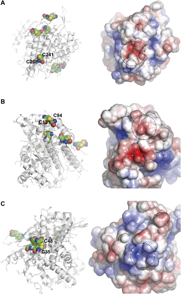
Fig. S4.
Highlighted view of cysteine residues in an inactive Ero1α mutant with the Cys94–Cys131 disulfide bond. (Left) Solvent-exposed cysteine residues are represented by spheres. (Right) Electrostatic potential of Ero1α, in which positively and negatively charged regions are shown in blue and red, respectively. Dotted circles highlight the regions around the indicated cysteine residues.

