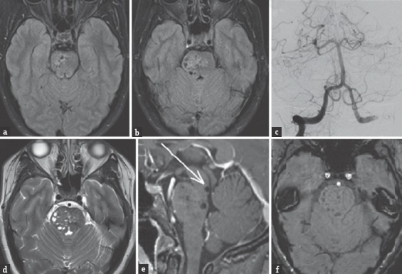
Figure 1.

(a and b) Axial fluid-attenuated inversion recovery (FLAIR) MRI performed at an outside hospital reveals multiple, oval-shaped hypointensities located within the right midbrain and pons. (c) Right vertebral artery injection diagnostic cerebral angiogram (Waters view) shows no cerebrovascular abnormalities. (d) Axial T2-weighted MRI with multiple cystic isointense (compared to cerebrospinal fluid) areas in midbrain and pons predominantly on right side. (e) Sagittal T1-weighted MRI with gadolinium contrast shows no associated enhancement. Note the close proximity of the patientfs dilated Virchow-Robin spaces (dVRS) to the cerebral aqueduct. This increases her risk for developing obstructive hydrocephalus if the dVRS continue to expand. (f) Axial susceptibility-weighted imaging (SWI) performed at our institution reveals no evidence of signal dropout to suggest hemorrhage or calcification
