
Figure 2.
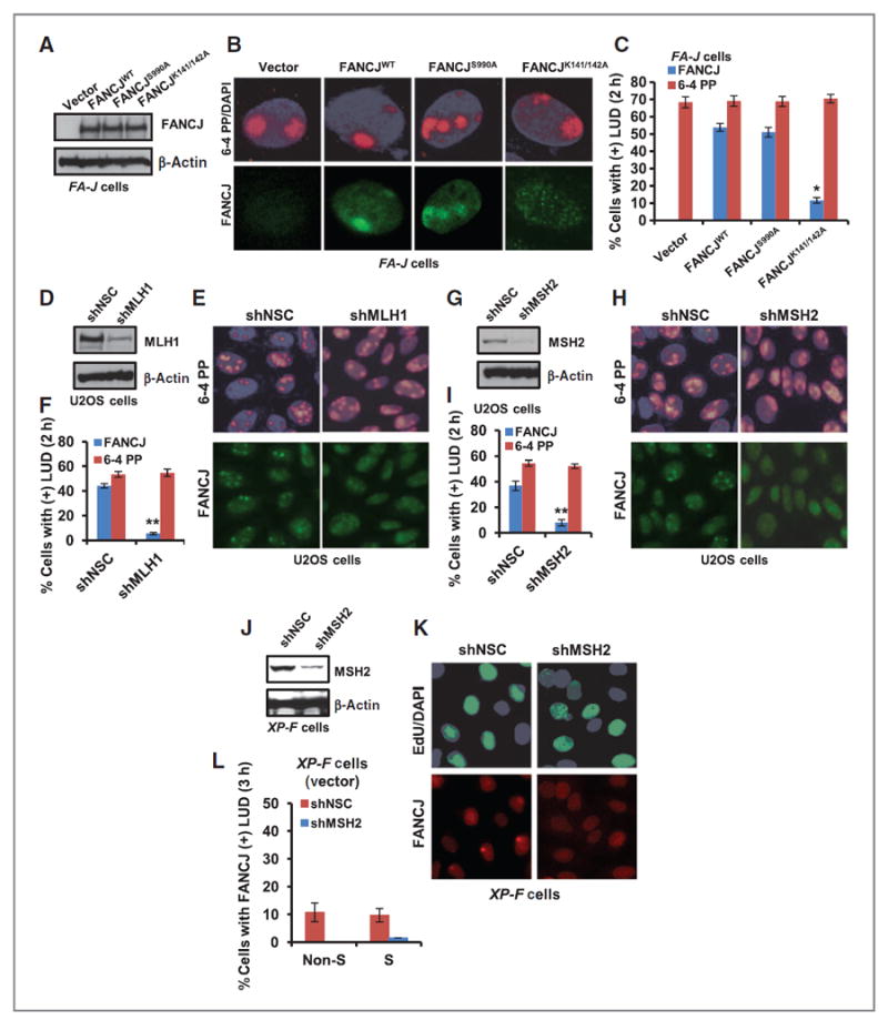
FANCJ recruitment to sites of LUDs is MMR dependent. A, FA-J cells were complemented with empty vector, FANCJWT, FANCJS990A, or FANCJK141/142A and analyzed by immunoblot. B and C, FA-J cells were UV irradiated through 5-μm micropore membrane filters, coimmunostained with the indicated Abs (B), and quantified for FA-J cells with FANCJ- and 6-4 PP–positive LUDs (C). D–F, U2OS cells containing shRNA vectors targeting MLH1 or NSC were analyzed by immunoblot (D) and UV irradiated through micropore filters (E) and quantified for cells with FANCJ- or 6-4 PP–positive LUDs (F). G–I, U2OS cells containing shRNA vectors targeting MSH2 or NSC were analyzed by immunoblot (G) and UV irradiated through micropore filters (H) and quantified for cells with FANCJ- or 6-4 PP–positive LUDs (I). J, XP-F cells complemented with empty vector were stably depleted of MSH2 versus NSC and analyzed by immunoblot. K, cells were treated as in E and processed for EdU incorporation and coimmunostained with the indicated Abs and quantified for cells with FANCJ-positive LUDs (L). Error bars represent the standard deviation of the mean of three independent experiments.