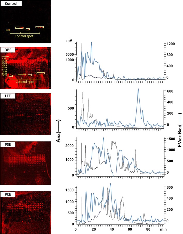
Fig. 2.

Binding signals of DBE-, LFE-, PSE-, and PCE-herbochips probed by GRP78. Left panels the images were visualized by Cy5-labeled streptavidin after the binding of b-GRP78 to DBE-, LFE-, PSE-, and PCE-herbochips, which were fabricated with extracts from Dioscorea bulbifera, Lasiosphaera fenzlii, Paeonia suffruticosa, and Polygonum cuspidatum, respectively. The spots in the control were 4, 10, 50, 250 ng/ml biotin in Optifix I, 1 μg/ml SA-Cy5 in Optifix II (positive control) and Optifix I (negative control) as those shown in Fig. 1 of Ref. [22]. Right panels the fluorescence intensity of the corresponding images were quantized by a scanner at 635 nm presented (back line) together with the original HPLC profile monitored at 254 nm (blue line). Positive signals indicated binding activity of the fractions to GRP78
