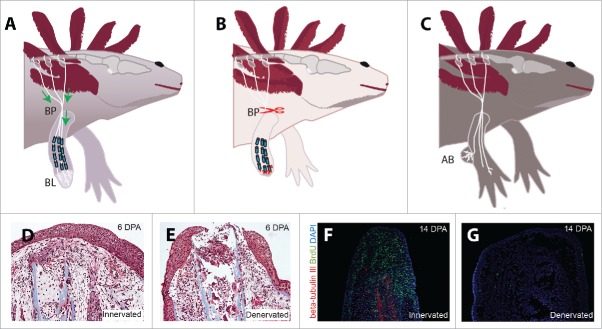
Figure 2.
(A) Diagram of the neurotrophic hypothesis, which states that nerve-secreted mitogens are transported from the dorsal root ganglia through the brachial plexus (BP) to support the proliferating blastema (BL). (B) Illustration of the inhibition hypothesis, in which denervation at the brachial plexus induces the release of inhibitory factors from Schwann Cells at the wound site. (C) Diagram of the accessory limb model, in which a peripheral nerve is deviated to a wound site to induce the formation of an accessory blastema (AB). (D) A control limb at 6 days post amputation (DPA). (E) A denervated limb at 6 DPA demonstrating extreme histolysis and inflammation. (F) The hyperinnervated regenerating blastema is highly proliferative at 14 DPA, as demonstrated by BrdU incorporation and staining. (G) Denervation eliminates beta-tubulin III staining of axons and substantially reduces the proliferative index of limbs at 14 DPA.

