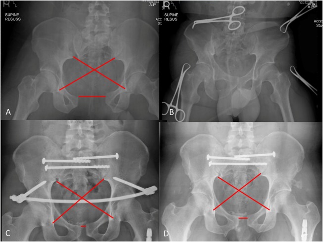
FIGURE 1.

AP-pelvis x-ray of implanted INFIX showing measurements taken to calculate PDI values and symphyseal widening. A, AP pelvis injury film, (B) after application of a binder, (C) after surgery with the INFIX and posterior fixation, and (D) after removal of INFIX. Editor's Note: A color image accompanies the online version of this article.
