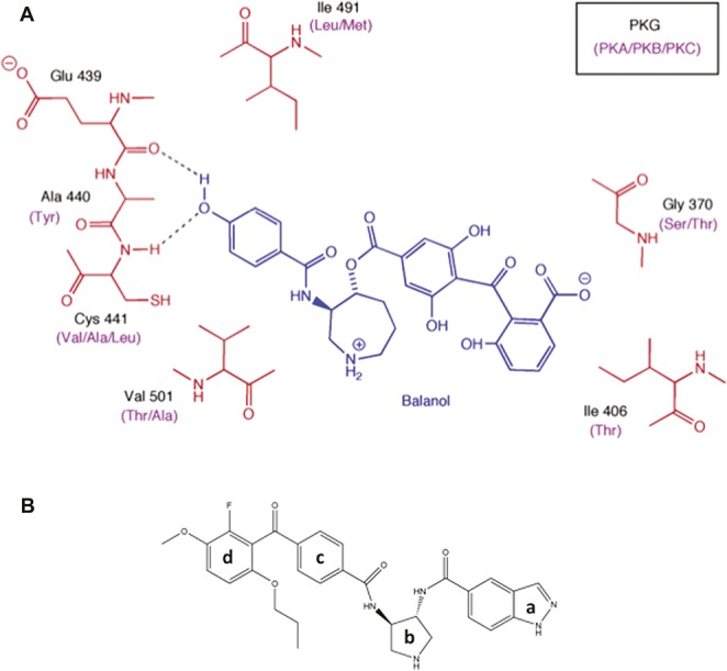
Figure 2.

Structural modeling of the PKG kinase domain with balanol. (A) 2D projection of balanol (blue) bound to PKG and PKA/B/C. The black labels indicate the amino acids in the binding site for PKG which differ from the corresponding amino acids (purple labels) in PKA, PKB, and PKC. (B) N46 consists of an indazole group (a) connected to a pyrrolidine ring (b) via an amide linkage. The pyrrolidine is linked to a benzophone ring (c) by an amide and the latter is attached via a ketone to a terminal benzophone ring (d).
