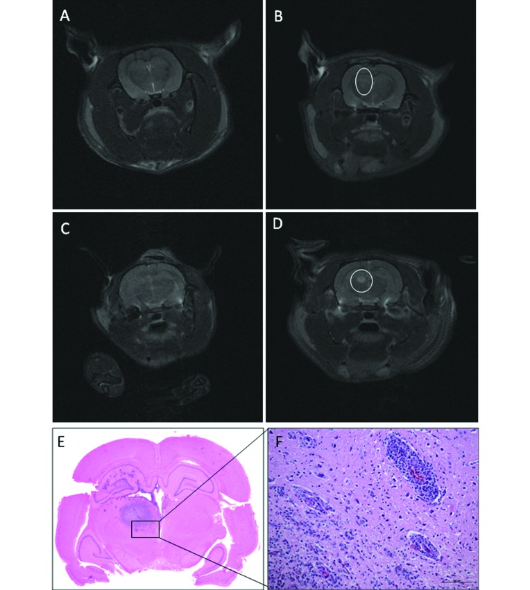
Figure 1.
Transverse T2-weighted MR images of Wistar rats that received C6-ATCC glioma cells. (A) Rat 12 on day 20: the needle tract is still identifiable, but no hyperintensity is present at the site of inoculation; the rat was judged as tumor negative. (B) Rat 4 on day 20: slight widening of the needle tract associated with hyperintense rim; the rat was judged as suspected (circle). (C) Rat 2 on day 20: no lesions detected. (D) Rat 2 on day 30: a well-defined, round, homogeneous hyperintensity is present in the caudate nucleus (circle). (E) Tumor mass. (F) Neovascularization and mononuclear cell infiltration present at the tumor periphery. Hematoxylin and eosin stain; bar, 100 µm.

