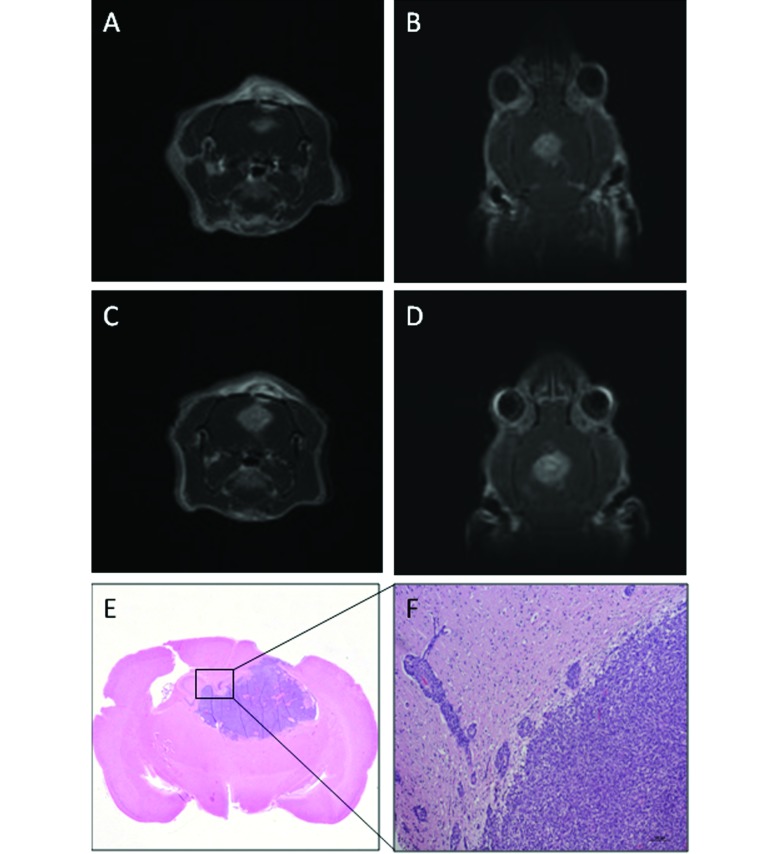
Figure 4.
Fischer rat (no. 29) that received F98 glioma cells. (A) Transverse and (B) dorsal T1-weighted MR images on day 9 show a well-defined hyperintense mass (volume, 86 mm3), which has small internal foci of increased hyperintensity. (C) Transverse and (D) dorsal T1-weighted MR images on day 11: tumor volume, 148 mm3. (E) Tumor mass. (F) Numerous neoplastic cells abutting adjacent blood vessels. Hematoxylin and eosin stain, bar, 100 µm.

