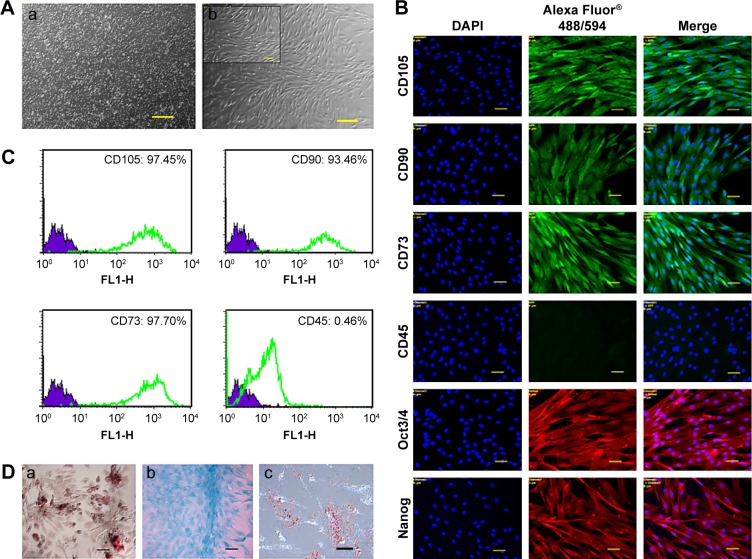
Figure 1.
Characterization of canine bone marrow-derived MSCs.
Notes: (A) Morphology of canine MSCs. (a) P0 after seeding and (b) P4 in confluence; scale bar: 100 µm (inset: 50 µm). (B) Immunofluorescence staining of MSC-specific surface markers (positive for CD105, CD90, and CD73 and negative for CD45) and pluripotency markers (Oct3/4, Nanog). Scale bar: 50 µm. (C) Immunophenotyping of canine MSCs at P4. Values represent the mean percentage of positively stained cells as analyzed by flow cytometry. (D) Potential of canine MSCs to differentiate into mesodermal lineages. (a) osteocytes (Alizarin Red staining), (b) chondrocytes (Alcian Blue staining), and (c) adipocytes (Oil Red O staining). Scale bar: 50 µm.
Abbreviations: CD, cluster of differentiation; DAPI, 4′,6-diamidino-2-phenylindole; MSC, mesenchymal stem cell; Nanog, unique homeobox transcription factor; Oct, octamer-binding transcription factor; P0/4, passage 0/4.

