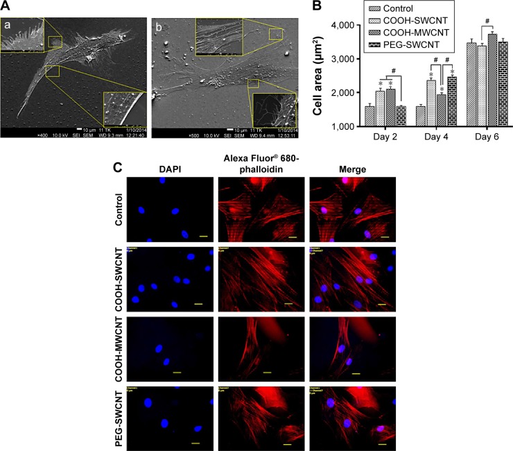
Figure 4.
Cellular behavior on CNT substrates.
Notes: (A) SEM observation of canine MSCs on (a) control and (b) CNT films after 4 days of culture. Insets show the magnified view of the cell boundary. In the control, cytoplasmic projections were less abundant and presence of lamellipodia at the edges of cells was visualized. In CNT film, a higher occurrence of nanosized filopodia suggests that CNT substrate exerts its influence on canine MSC morphology. (B) Cell spreading area on control and CNT films at different time intervals. The symbols * and # indicate significance (P<0.05) with respect to control and between the films, respectively, on a particular day. The results are the mean ± standard error of the mean of triplicate experiments (n=30). (C) Alexa Fluor 680-conjugated phalloidin-labeled F-actin (red), DAPI nuclear staining (blue), and merged fluorescence images of immunostained cellular components of canine MSCs cultured on control and different CNT films on Day 4. Scale bars: 20 µm.
Abbreviations: CNT, carbon nanotube; DAPI, 4′,6-diamidino-2-phenylindole; MSC, mesenchymal stem cell; MWCNT, multiwalled CNT; PEG, polyethylene glycol; SEM, scanning electron microscopy; SWCNT, single-walled CNT.

