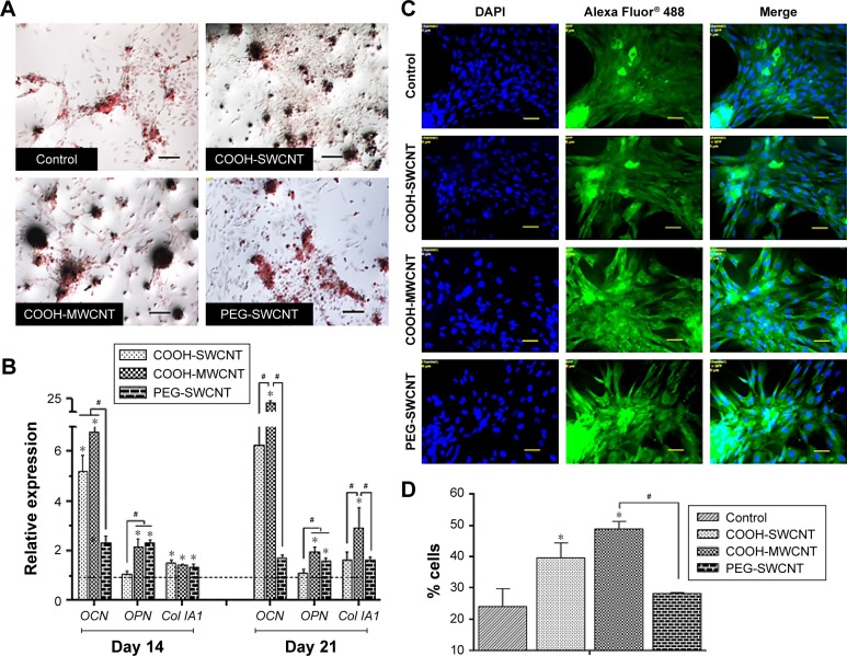
Figure 6.
Induced osteogenic differentiation of canine MSCs on CNT films.
Notes: (A) Representative images of Alizarin Red staining after 21 days of osteogenic differentiation in control and different CNT films. The size and darkness of mineralized nodules were higher on CNT substrates compared to control. Scale bars: 100 µm. (B) Relative expression of bone marker genes of canine MSCs differentiated on control and different CNT films. Results normalized to GAPDH relative to control. Dashed line indicates values of target genes in control conditions. The symbols * and # indicate significance (P<0.05) with respect to control and between the films, respectively, for a target gene. The results are the mean ± standard error of the mean of the triplicate experiments. (C) Immunofluorescence of osteocalcin-positive cells (green), DAPI nuclear staining (blue), and merged fluorescence images on control and CNT films after 21 days. Scale bars: 50 µm. (D) Flow cytometry assay of osteocalcin-positive cells (percentage) after 21 days of osteogenic differentiation on control and different CNT films. The symbols * and # indicate significance (P<0.05) with respect to control and between the films, respectively. The results are the mean ± standard error of the mean of the triplicate experiments.
Abbreviations: CNT, carbon nanotube; DAPI, 4′,6-diamidino-2-phenylindole; GAPDH, glyceraldehyde 3-phosphate dehydrogenase; MSC, mesenchymal stem cell; MWCNT, multiwalled CNT; PEG, polyethylene glycol; SWCNT, single-walled CNT.

