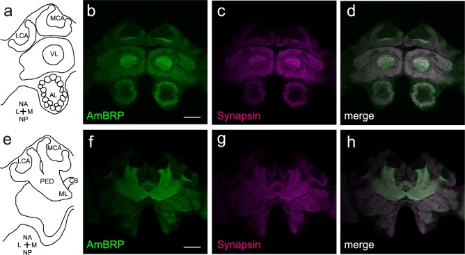
Fig 2. Distribution of AmBRP and Synapsin in the honeybee central brain.
Optical sections of a honeybee central brain incubated with the anti-BRPlast200 and anti-SYNORF1 to visualize the presynaptic proteins AmBRP and Synapsin. a Longitudinal section through a schematic honeybee brain showing ventrally located regions (nomenclature after Ito et al. (2014) [34]). b-d Distribution of BRPlast200 signals (b) and anti-SYNORF1 signals (c) in ventrally located brain regions of a 29-day-old bee. Both antibodies show staining in all brain regions with almost similar distribution (d). Prominent stained regions are the vertical lobes and the antennal lobes. e Longitudinal section through a schematic honeybee brain showing dorsally located regions (nomenclature after Ito et al. (2014) [34]).f-h Distribution of BRPlast200 signals (f) and anti-SYNORF1 signals (g) in dorsally located brain regions of a 29-day-old bee. Both antibodies show staining in all brain regions with similar distribution (h) Prominent stained regions are the peduncles, especially in the AmBRP staining. LCA, lateral calyx; MCA, medial calyx; VL, vertical lobe; PED, peduncle; ML, medial lobe; CB, central body; AL, antennal lobe; NA, neuraxis anterior; M, medial; NP, neuraxis posterior; L, lateral. Scale bars: 200 μm for b-d and f-h.

