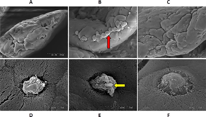
Fig 3. Ultrastructure of mucosal epithelial cells of ileum under a scanning electron microscope.
(A) and (D): control group; (B) and (E): water-control group; (C) and (F): IMO-treated group. A, B, C: ×1200; D, E, F: ×10000. Long arrow: secretory granules at the opening of the mucosal glands; short arrow: bacilli on goblet cells.

