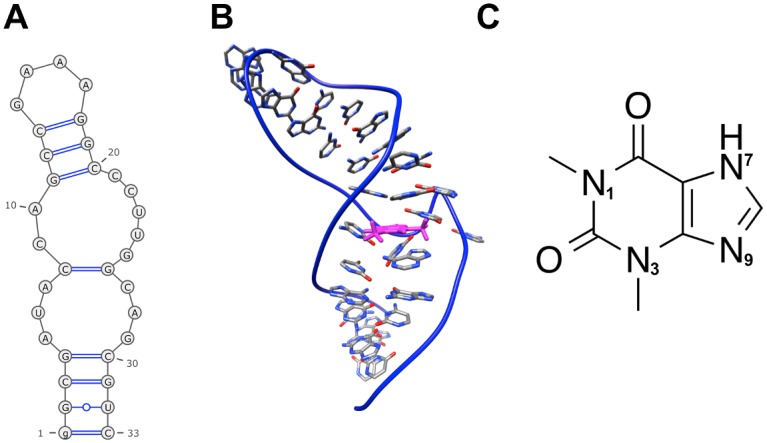
Fig 2. Solution NMR structure of theophylline aptamer in the bound state.

(A) Secondary structure. (B) Tertiary structure. The bound theophylline molecule is colored magenta. (C) Theophylline structure. Depicted RNA structures are based on the first conformer of PDB entry 1EHT. Secondary structure diagram was generated by the RNApdbee webserver[88].
