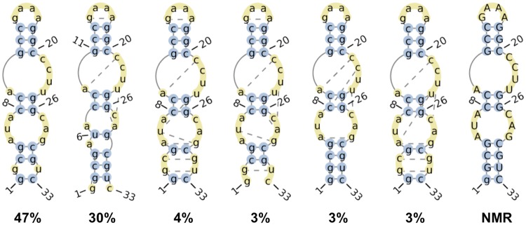
Fig 7. Six most frequently observed RNA secondary structures in the absence of bound theophylline.
Numbers denote proportions of all analyzed simulation snapshots in which the RNA adopts the depicted secondary structures. Bases that participate in canonical and non-canonical base pairing interactions are depicted in blue circles and yellow circles connected by dotted lines, respectively. Unpaired bases are depicted in unconnected yellow circles. The secondary structure of the bound-state NMR structure is shown on the far right. Secondary structure diagrams were generated by the RNApdbee webserver[88].

