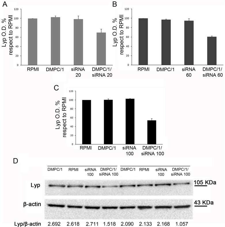
Fig 4. Lyp protein levels in Jurkat T cells 48 hours after the beginning of O/N transfection with different doses of siRNA s/a in DMPC/1/siRNA lipoplexes.
Representative duplicate experiment among all replicas in S4 Fig. (A) Lyp expression in Jurkat T cells cultured in RPMI or after O/N transfection with DMPC/1, 20 pmols of siRNA and 20 pmols of siRNA in DMPC/1/siRNA lipoplexes (DMPC/1/siRNA20). 20 pmols of siRNA complexed with DMPC/1 resulted in a 31% reduction of Lyp expression. (B) Same experiment as in A using 60 pmols of siRNA complexed with DMPC/1 (DMPC/1/siRNA60). 39% reduction of Lyp expression was obtained. (C) Same experiment as in A using 100 pmols of siRNA complexed with DMPC/1 lipoplexes (DMPC/1/siRNA100). 47% reduction of Lyp expression was obtained. (D) Representative WB image within all experimental groups is shown. Under each blot Lyp O.D. values for every treatment are normalized over the corresponding β-actin values. All percentages were expressed relatively to untransfected cells (RPMI) that is considered the 100% of basal Lyp expression. Graphs A, B, C show the mean values and their standard deviations. In all experimental conditions (vide infra), a decrease in Lyp protein level was not observed in cells treated with the liposome alone or, more importantly, in cells treated with the correspondent dose of the siRNA s/a free molecule.

