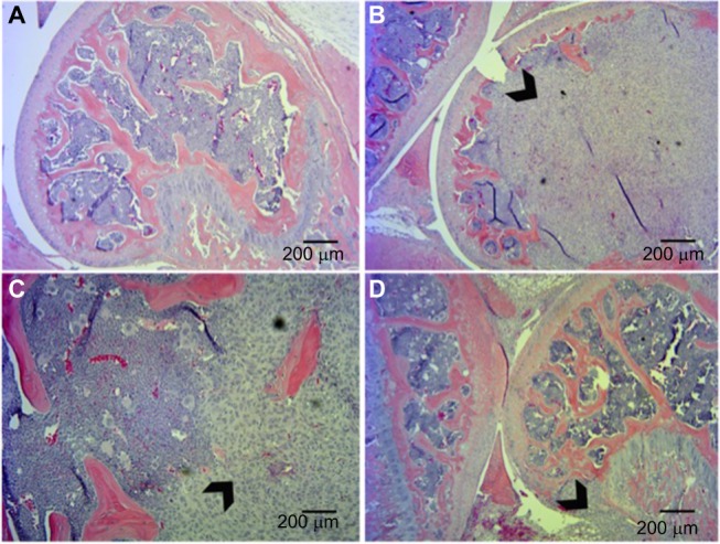
Figure 4.

Histological analysis of tumor xenografts in bone represented by hematoxylin and eosin staining.
Notes: (A) Sham-injected mice. Tumor-injected mice show extensive invasion of xenograft into the femur with destruction of growth plate (B, D) and in some cases breaching of the periosteum (C). Arrowheads indicate tumor tissue.
