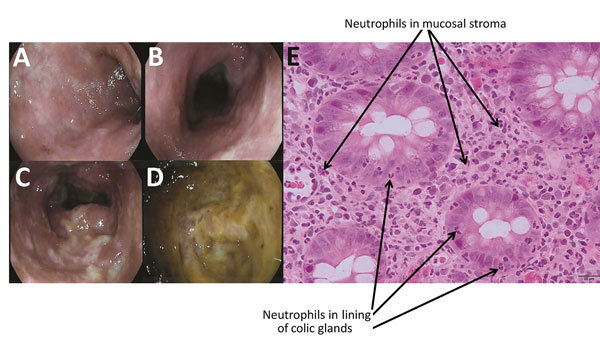
Figure.

Endoscopic imagery of the distal sigmoid colon (A), proximal sigmoid colon (B), descending colon (C), and base of cecum (D), revealing diffuse colitis with mucosal erythema, edema, and mucopurulent exudate without ulceration. Colonic biopsy (E) demonstrating neutrophilic infiltrates (indicated with arrows) in the epithelial lining of the colic glands and the mucosal stroma compatible with mild active colitis without signs of chronicity.
