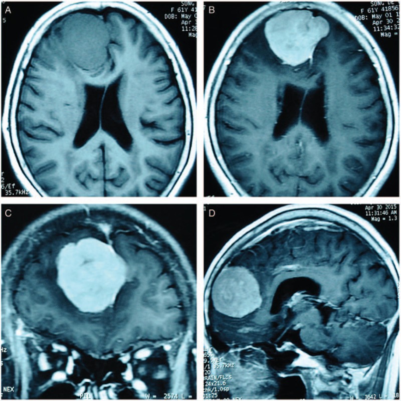
Figure 1.

Preoperative plain and contrast-enhanced MRI of the head. The tumor is approximately 5.7 × 5.0 × 5.0 cm3 and has a characteristic “dural-tail” sign in the contrast-enhanced imaging. A is the axial view of the T1-weighted image, which shows an isointensity mass. The tumor is homogenously contrast-enhanced and shown in axial (B), coronal (C), and sagittal (D) views. MRI = magnetic resonance imaging.
