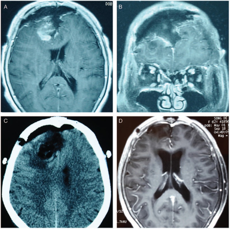
Figure 2.

Radiological findings of the patient during the postoperative and follow-up periods. The axial (A) and coronal (B) views of the contrast-enhanced MRI on postoperative day 3 revealed the radical removal of the tumor and no sign of intracranial hemorrhage or brain swelling. Emergency brain CT (postoperative day 4) (C) failed to reveal any abnormal findings that would indicate brain swelling, intracranial hemorrhage, or cerebral infarction. A follow-up MRI 5 months postoperation (D) showed a clear operative field. CT = computed tomography, MRI = magnetic resonance imaging.
