
Figure 1.
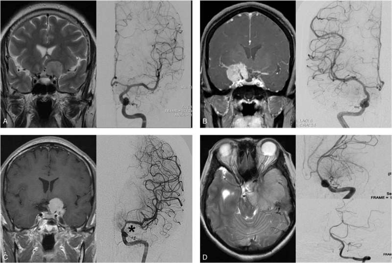
Representative radiographic images according to Goel's classification. Type A; Coronal T2-weighted magnetic resonance imaging (MRI) (left) and angiography (right) show just a medially displaced internal cerebral artery (ICA). Type B; Coronal T1-contrast enhanced MRI (left) and angiography (right) shows encased but not narrowed ICA by the tumor. Type C; Coronal T1-contrast enhanced MRI (left) and angiography (right) reveals encased and narrowed ICA by the tumor (asterisk). Type D; axial T2-weighted MRI (left) and angiography (right-upper; ICA, right-lower; vertebral artery) shows displaced ICA and basilar artery by the tumor. ICA = internal carotid artery, MRI = magnetic resonance imaging.