
Figure 1.
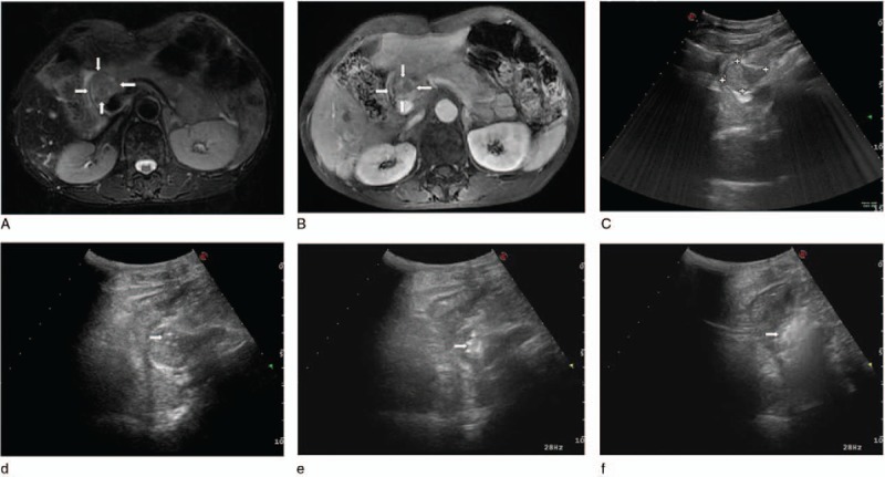
(A) Preoperative axial T2-weighted (white arrows), (B) substance phase MRI image of the abdomen showed a mass adjacent to the hepatic portal vein, pancreas, and stomach (white arrows). (C) Axial gray-scale US image of the retroperitoneal region showed the mild hyperechoic area. (D) Intraoperative sonogram indicated the arranging needle method of using 2 laser fibers parallelly ablating the tumor under US guidance. Initial postoperative immediate US showed local enhancement (arrowhead) (E) and global enhancement (arrowhead) (F) of the lesions. MRI = magnetic resonance imaging, US = ultrasound.