
Figure 2.
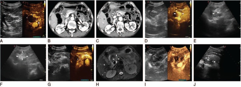
(A) Comparing to conventional ultrasound, the CEUS of the next day showed a little remanent tumor (arrowhead), and there was no detectable enhancement in the necrosis area in the center of the whole mass. Coronal contrast-enhanced CT images acquired 5 days after initial ablation showed lower tumor low signal intensity (arrowhead) (B) and upper mass intermediate high signal intensity (arrowhead) (C) in substance phase, and blood vessel was not injured. (D) One week later, these residuary small nodules appear fusion during CEUS image (arrowhead). (E) Two laser fibers were parallelly accurately inserted into the lesion with US guidance (arrowheads). (F) Axial gray-scale US image showed increased echogenicity covering the whole mass (white arrows). On the next day of the 2nd ablation, postoperative CEUS images showed they were still remanent (G). At the corresponding MRI, it also showed this in left of tumor (arrowheads) (H). After the CEUS positioning the remanent tumor (I), the 3rd laser ablation was performed using 2 laser fibers (arrowheads) (J). CEUS = contrast-enhanced ultrasound, CT = computed tomography, MRI = magnetic resonance imaging, US = ultrasound.