
Figure 3.
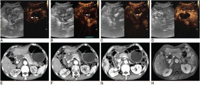
Because remanent tumor was detectable under CEUS guidance 3 days after 3 ablations (arrowheads) (A), the single needle was accurately inserted into the residual lesion (arrowheads) (B), showing local enhancement (arrowheads) (C) and no contrast agents filled (D). Substance phase of CT obtained 3 days after US-guided percutaneous LA showed complete necrosis of the tumor (white arrows) (including upper [white arrows] (E), mid [white arrows] (F), and lower tumor [white arrows] (G) of it), and 1 month later substance phase MR image had corresponding results (H). CEUS = contrast-enhanced ultrasound, CT = computed tomography, LA = laser ablation, MR = magnetic resonance, US = ultrasound.