Figure 2.

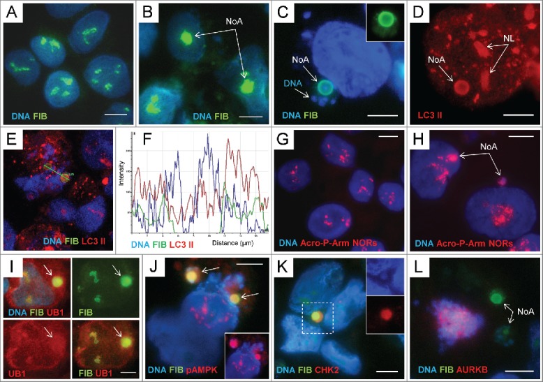
The fibrillarin-positive nucleolar aggresomes include damaged rDNA, autophagy markers and are released into cytoplasm accompanied by heterochromatin: (A) – immunostaining for FIBRILLARIN (FIB) and counterstaining with DAPI in non-treated PA1 cells; (B) the same staining of ETO-treated cells. The round nucleolar FIB-positive aggregates are designated as the nucleolar aggresomes NoA; (C, D) the perinuclear ring-shaped NoA colocalising FIB with LC3-II and DAPI (inside) and surrounded by DAPI-positive granular chromatin. The nucleoli (NL) designated on (D) are also positive for LC3-II; (E, F) confocal section through the interior of 2 NoA bodies and the plot of fluorescence intensity in the region of interest showing colocalization of all 3 components in the sample stained with 3 fluorochromes for FIB, LC3-II, and DNA (note a DAPI-peak inside); (G) FISH with the probe Acro P-arm-NORs counterstained with DAPI in NT PA1 cells; (H) the same reaction applied on ETO-treated cells where NORs are revealed both in intranuclear and perinuclear NoA (arrowed); (I) accumulation of UBIQUITIN 1(UB1) in the perinuclear FIB-positive NoA; (J) Colocalization of FIB with pAMPK in 2 NoAs (imaged in RGB optical filter); on insert, the same image in a red channel for pAMPK; (K) a cytoplasmic NoA composed of the largely colocalising FIB, CHK2, and the DNA containing DAPI-stained core (on inserts); (L) Fragment of the mitotic cell (tripolar mitosis) highlighted by DAPI and AURKB containing 2 NoAs in the cytoplasm. Bars = 10 µm.
