
Fig. 10.
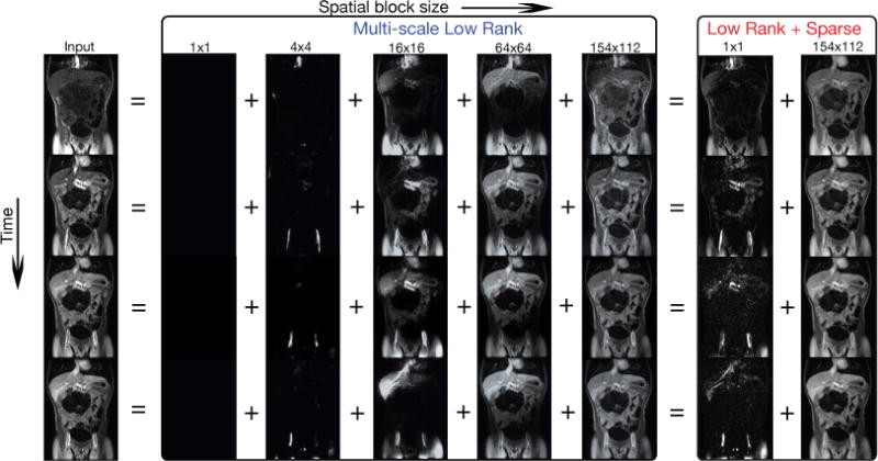
Multi-scale low rank versus low rank + sparse decomposition on a dynamic contrast enhanced magnetic resonance image series. For the multi-scale result, small contrast dynamics in vessels are captured in 4 × 4 blocks while contrast dynamics in the liver are captured in 16 × 16 blocks. The biggest block size captures the static tissues and interestingly the respiratory motion. In contrast, the low rank + sparse modeling could only provide a coarse separation of dynamics and static tissue, which result in neither truly sparse nor truly low rank components.