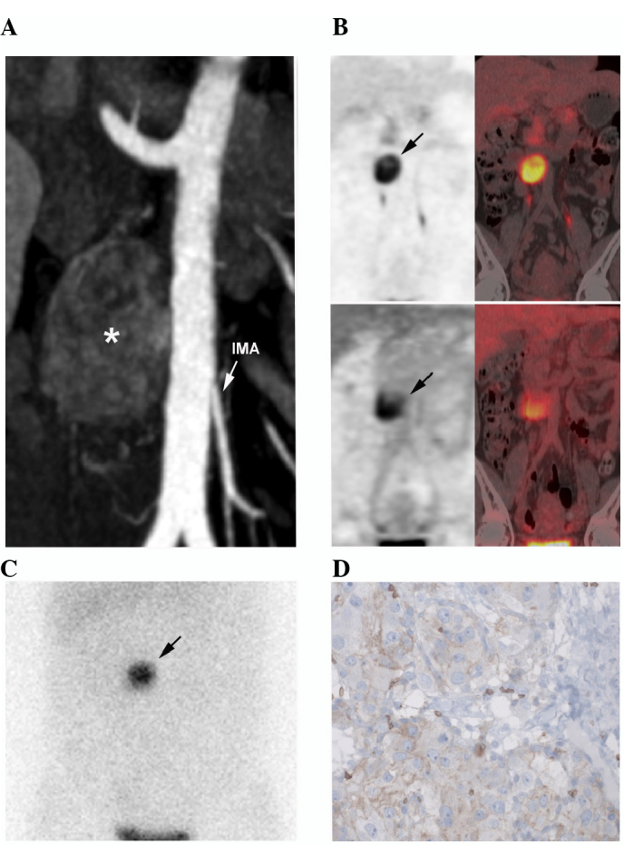
Figure 1.

Imaging and pathological features of the OZ-PGL. (A) Contrast-enhanced CT (arterial phase) showing a 40-mm hypervascular and heterogeneous left para-aortic mass located at the level of the IMA (asterisk). (B) 18F-FDOPA (upper image) and 18F-FDG PET/CT (lower image) imaging showing a single tumor. (C) Iodine-123-metaiodobenzylguanidine scintigraphy also positively located the mass (planar anterior view). (D) Immunohistochemical analysis of the tumor demonstrated positive glucose transporter-1 immunostaining (~10%). CT, computed tomography; IMA, inferior mesenteric artery; 18F-FDOPA, 18fluorine-L-dihydroxyphenylalanine; 18F-FDG, 18F-fluorodeoxyglucose; PET, positron emission tomography.
