Figure 2.
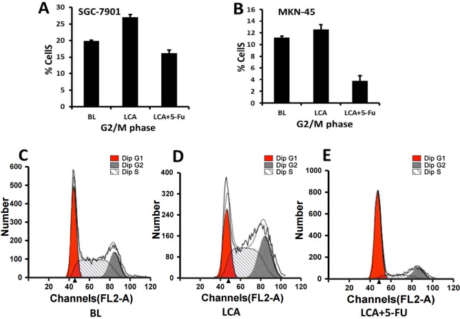
Effects of LCA on the cell cycle progression of gastric cancer cells. (A) SGC-7901 and (B) MKN-45 cell lines were exposed to BL, LCA and LCA combined with 5-FU for 48 h. The cell cycle distributions of SGC7901 cells when exposed to (C) blank control, (D) LCA (which induced growth inhibition associated with G2/M arrest) and (E) LCA combined with 5-FU (which induced G0/G1 arrest more markedly) for 48 h are presented. All assays were performed in triplicate. BL, blank control; LCA, licochalcone A; 5-FU, 5-fluorouracil.

