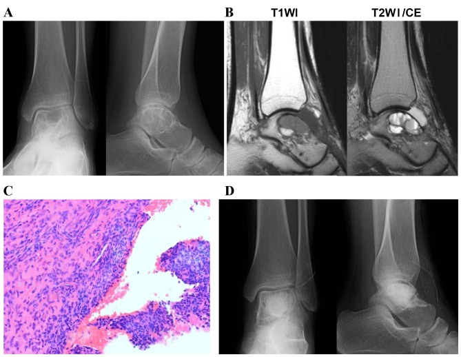
Figure 1.
A case of primary ABC that occurred in the talus and recurred following curettage. (A) Radiography revealed a multiloculated osteolytic lesion close to the articular surface of the talus. (B) MRI revealed multiple cystic lesions divided by thin septa. (C) A histological tissue section obtained from the curettage specimen exhibited collagenous tissue with spindle-shaped cells and sporadic multinucleated giant cells. Original magnification, ×200. (D) Postoperative radiography revealed the artificial bone graft in the lesion. T1WI, T1-weighted image; T2WI, T2-weighted image; ABC, aneurysmal bone cyst; MRI, magnetic resonance imaging.

