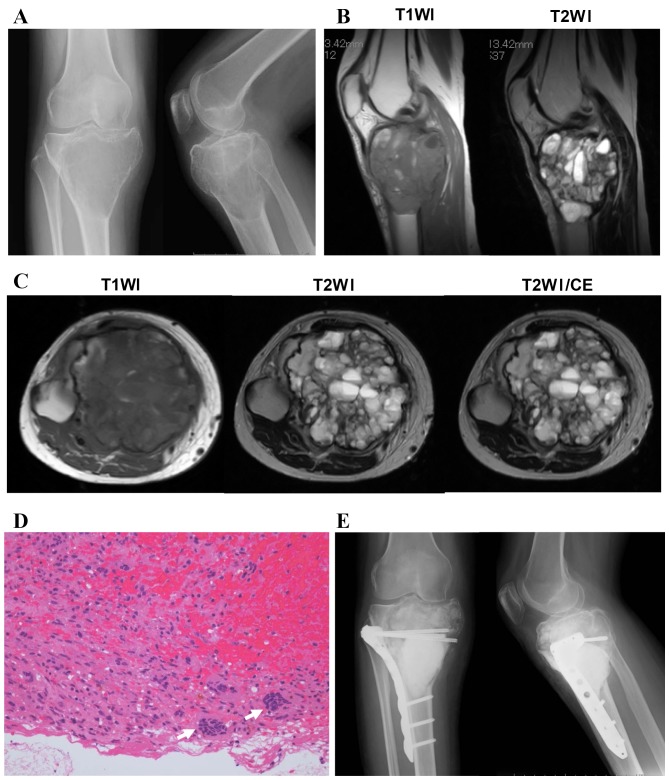
Figure 2.
A case of secondary ABC with a giant cell tumor. (A) Radiography demonstrated an eccentric multiloculated osteolytic lesion in the metaphysis of the proximal tibia. (B) Sagittal and (C) axial MRI revealed multiple cystic lesions with a soft tissue mass. (D) A histological tissue biopsy section exhibited stromal cells with round nuclei and multinucleated giant cells (arrows). Original magnification, ×200. (E) Following the pathological diagnosis of a giant cell tumor, curettage and PMMA cementing, supported with a locking plate, was performed. T1WI, T1-weighted image; T2WI, T2-weighted image; CE, contrast-enhanced imaging; ABC, aneurysmal bone cyst; MRI, magnetic resonance imaging; PMMA, polymethyl methacrylate.

