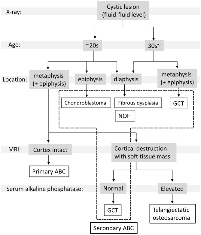
Figure 3.
A simplified flowchart to facilitate discrimination between ABC and other types of bone tumors during diagnosis. Patients with bone tumors exhibited multiple cystic lesions in X-rays are first stratified by age. In patients ≥30 years old, the occurrence of a secondary ABC following a fibrous dysplasia or giant cell tumor should be considered. Subsequently, based on the location of the initial lesion, a primary differential diagnosis may be established. In patients <30 years old with a lesion in the metaphysis, cortex destruction must be evaluated using MRI. If the cortex is intact, the most likely diagnosis is primary ABC. If the cortex is degenerating and a soft tissue mass is present, an aggressive bone tumor, including a giant cell tumor or telangiectatic osteosarcoma, must be considered. In cases with elevated serum alkaline phosphatase levels, a telangiectatic osteosarcoma is possible. ABC, aneurysmal bone cyst; MRI, magnetic resonance imaging; GCT, Giant cell tumor; NOF, Non ossifying fibroma.

