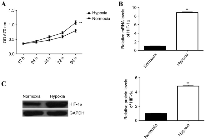
Figure 1.
(A) An MTT assay was performed to determine the proliferation of EPCs cultured under hypoxia or normoxia for 12, 24, 48, 72 and 96 h. (B) Reverse transcription-quantitative polymerase chain reaction and (C) western blotting were performed to measure HIF-1α mRNA and protein expression, respectively, in EPCs cultured under hypoxia or normoxia. GAPDH was used as an internal reference. **P<0.01 vs. normoxia. HIF-1α, hypoxia-inducible factor 1α; OD, optical density; EPCs, endothelial progenitor cells.

