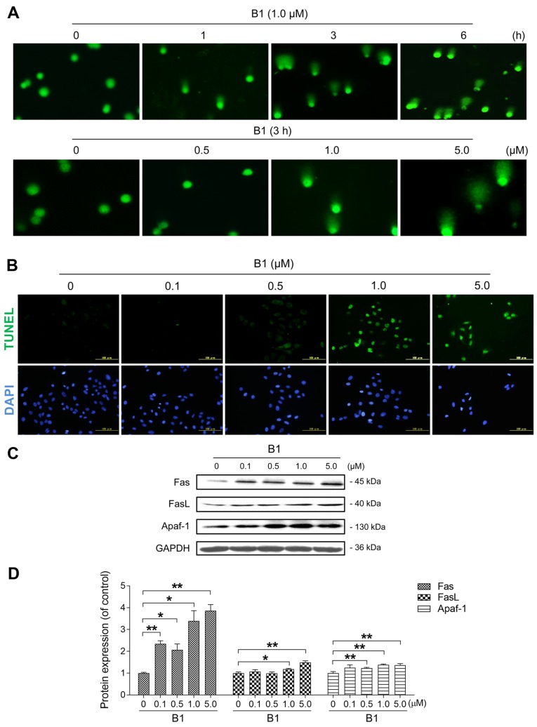
Figure 2.
B1 treatment induces DNA damage and apoptosis in A549 cells. (A) Comet images of DNA double-strand breaks were obtained in cells treated with 1.0 µM B1 at various time intervals (×100), or with the indicated concentrations of B1 for 3 h (×200). (B) A549 cells were exposed to different concentrations of B1 for 24 h, and apoptosis was determined with terminal deoxynucleotidyltransferase-mediated dUTP nick end labelling (TUNEL) staining, followed by fluorescence detection. (C) Western blot analysis was used to detect apoptosis regulatory proteins in A549 cells treated with various concentrations of B1 for 48 h. (D) Protein expression of Fas, FasL and Apaf-1 after normalization against GAPDH. The data are expressed as the mean ± SE for three independent experiments with similar results. *P<0.05 and **P<0.01 vs. controls.

