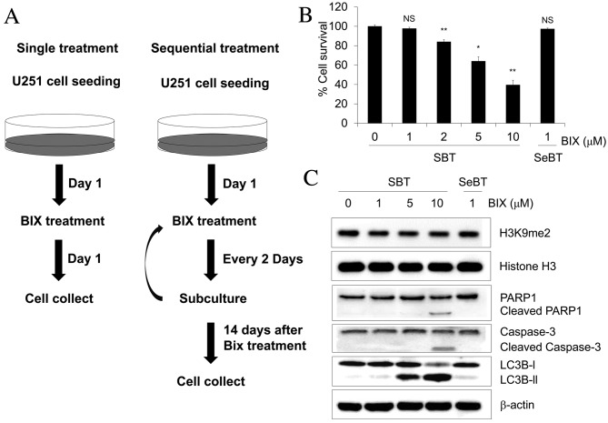
Figure 1.
Cell survival effect of Bix treatment following the indicated dosing schedule in human U251 glioblastoma cells. (A) Experimental scheme for Bix treatment. (B) The cells were treated with 1, 2, 5 or 10 µM Bix and the methyl thiazolyl tetrazolium assay was performed. (C) Levels of dimethylated H3K9 and apoptosis/autophagy-associated genes investigated by western blot analysis. Relative optical densities of dimethylated H3K9 level were normalized to total histone H3 (1, 0.88±0.01, 0.80±0.05, 0.91±0.03, 0.92±0.1, respectively, mean ± standard error of the mean). β-actin was used as the loading control. Western blotting was performed at the indicated dosages (0, 1, 5 and 10 µM). Bix, BIX01294; SBT, single treatment of Bix; SeBT, sequential treatment of Bix; PARP, poly (ADP-ribose) polymerase. P-values were calculated using the Student's t-test. *P<0.01 vs. the control; **P<0.001 vs. the control; N.S. no significance.

