
Figure 1.
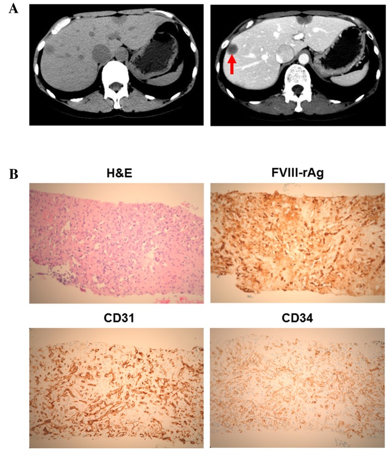
Clinicopathological features of the present case of hepatic epithelioid hemangioendothelioma. (A) Computed tomography revealed hypodense tumors in the two lobes (left panel). In the portal phase, the center of the tumor was enhanced in the right lobe (right panel; red arrow). (B) In the H&E-stained section, tumor cells were observed to form intracytoplasmic lumina, and solitary, alveolar and single-file patterns were detected. Immunohistochemical staining demonstrated that the tumor cells were positive for FVIII-rAg, CD31 and CD34. FVIII-rAg, factor VIII-associated antigen; CD31, platelet endothelial cell adhesion molecule 1; CD34, human hematopoietic progenitor cell antigen; H&E, hematoxylin and eosin.