
Figure 2.
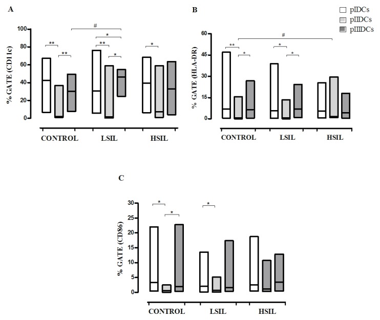
Phenotypic profiles of monocyte-derived pIDCs, pIIIDCs and pIIDCs. PBMCs were stimulated with GM-CSF, IL-4 and TNF-α to generate pIDCs. The expression of co-stimulatory molecules and surface markers (A) CD11c, (B) HLA-DR and (C) CD86 and was evaluated by flow cytometry to evaluate the effect of different protocols of differentiation on the maturation profile. PBMCs were stimulated with GM-CSF, IL-4, TNF-α and activated lymphocytes in the absence of non-adherent cells to generate pIIDCs. PBMCs were stimulated with GM-CSF, IL-4, TNF-α and activated lymphocytes in the presence of non-adherent cells to generate pIIIDCs. Data are presented as the median and range (Mann-Whitney and Kruskal-Wallis tests). #P<0.05 (comparison between patient groups); *P<0.05, **P<0.01 (comparison between protocols). Control, healthy patients. LSIL, low-grade squamous intraepithelial lesion; HSIL, high-grade squamous intraepithelial lesion; PBMC, peripheral blood mononuclear cell; pI–III, protocol I–III; CD, cluster of differentiation; DC, dendritic cell; HLA-DR, human leukocyte antigen-antigen D related; GM-CSF, granulocyte-macrophage colony-stimulating factor; IL, interleukin; TNF, tumor necrosis factor.