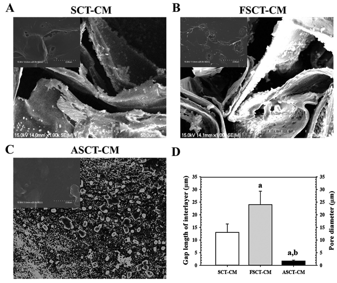
Figure 1.
Scanning electron microscopy (SEM) images of the three Styela clava tunic-cellulose membranes (SCT-CMs). The ultrastructure of the fracture surface of (A) SCT-CM, (B) FSCT-CM and (C) ASCT-CM was observed by SEM at ×1,000 magnification as described in the Materials and methods. The images in upper left corner represent the ultrastructure of the surface. (D) The gap length of interlayer in SCT-CM and FSCT-CM and pore size (µm) of each ASCT-CM was measured was measured using Leica Application Suite as described in the Materials and methods. Three to five films per group were assayed in duplicate by SEM. aP<0.05 compared to SCT-CM; bP<0.05 compared to FSCT-CM. FSCT-CM, freeze-dried SCT-CM; ASCT-CM, sodium alginate-supplemented SCT-CM.

