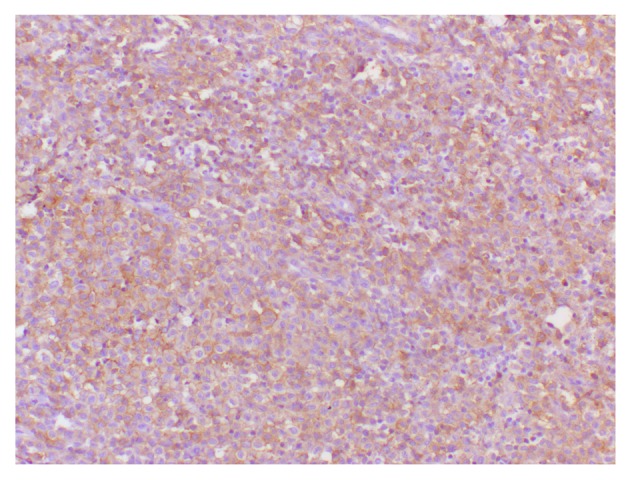
Figure 3.

Immunohistochemical observations demonstrated histiocytic cells that were positive for CD1a expression, which was consistent for cells of Langerhans' origin. The image depicts an immunoperoxidase staining technique (original magnification, ×400).
