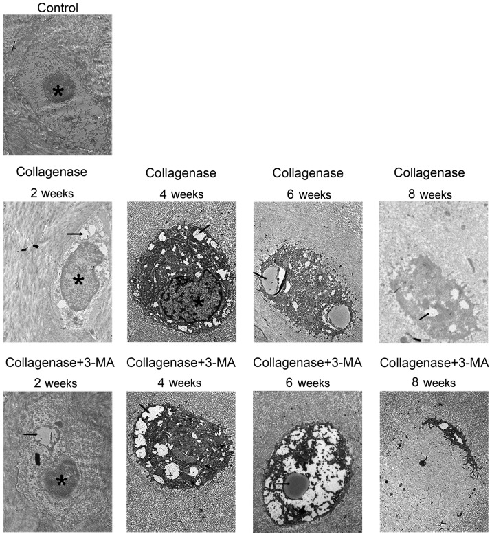
Figure 5.
Representative images of transmission electron microscopy from control, collagenase-injected and 3-methyladenine (3-MA)-treated rabbits. Autophagosomes and chondrocyte degeneration were observed. Autophagosomes in images are marked with black arrows, the nuclei are marked with an asterisks (*). Scale bar, 1 μm.

