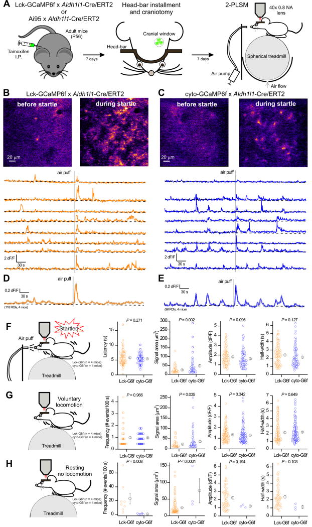
Figure 5. Comparison of in vivo astrocyte calcium signals measured with Lck-GCaMP6f and cyto-GCaMP6f driven by Aldh1l1-Cre/ERT2 mice.
A. Schematic drawings showing the workflow for in vivo 2PLSM in head-fixed, awake mice. In brief, after the mice were injected with tamoxifen for 7 days, a lightweight metal head bar was glued to their skull and a 3 mm cranial window was made above the visual cortex. Mice were then head-fixed onto a spherical treadmill where they were free to rest or run. A 40 x objective lens as part of a 2-photon laser scanning imaging microscope was positioned above the cranial window. An air pump outside the microscope enclosure was used to generate an unexpected air puff, which evoked startle response. B & C. Top: Representative pseudo-colored images showing the fluorescence increase of membrane-tethered Lck-GCaMP6f (B) and cyto-GCaMP6f (C) in astrocytes of mouse visual cortex before and during startle. Bottom: representative ΔF/F traces from 8 randomly selected ROIs (10 μm2 each) in Lck-GCaMP6f x Aldh1l1-Cre/ERT2 mice (orange) and cyto-GCaMP6f x Aldh1l1-Cre/ERT2 (blue). The gray vertical line indicates the air puff. The trend for the baseline is shown in black. D. The average F/F trace of 118 ROIs from four Lck-GCaMP6f x Aldh1l1-Cre/ERT2 mice (s.e.m. shown with gray lines for every 5th time point). E. The average F/F trace of 96 ROIs from four cyto-GCaMP6f x Aldh1l1-Cre/ERT2 mice (s.e.m. shown with gray lines for every 5th time point). F. Comparison of calcium signals detected by Lck-GCaMP6f (113 events, n = 4 mice) and cyto-GCaMP6f (94 events, n = 4 mice) during startle. G. Comparison of calcium signals detected by Lck-GCaMP6f and cyto-GCaMP6f during voluntary locomotion (without air puff). H. Comparisons of calcium signals detected by Lck-GCaMP6f and cyto-GCaMP6f during the resting phase (without air puff and locomotion).

