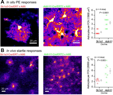
Figure 6. Aldh1l1-Cre/ERT2 mice permit high-density imaging of calcium signals in vivo and in brain slices.
A. Representative images and average data of 10 μM PE-evoked astrocyte calcium signals in visual cortex brain slices when cyto-GCaMP6f was driven by Slc1a3-Cre/ERT2 or by Aldh1l1-Cre/ERT2. B. As in A, but for in vivo startle-evoked response in the visual cortex. In both cases, many more astrocytes were detected when cyto-GCaMP6f was driven by the Aldh1l1-Cre/ERT2 mouse.

