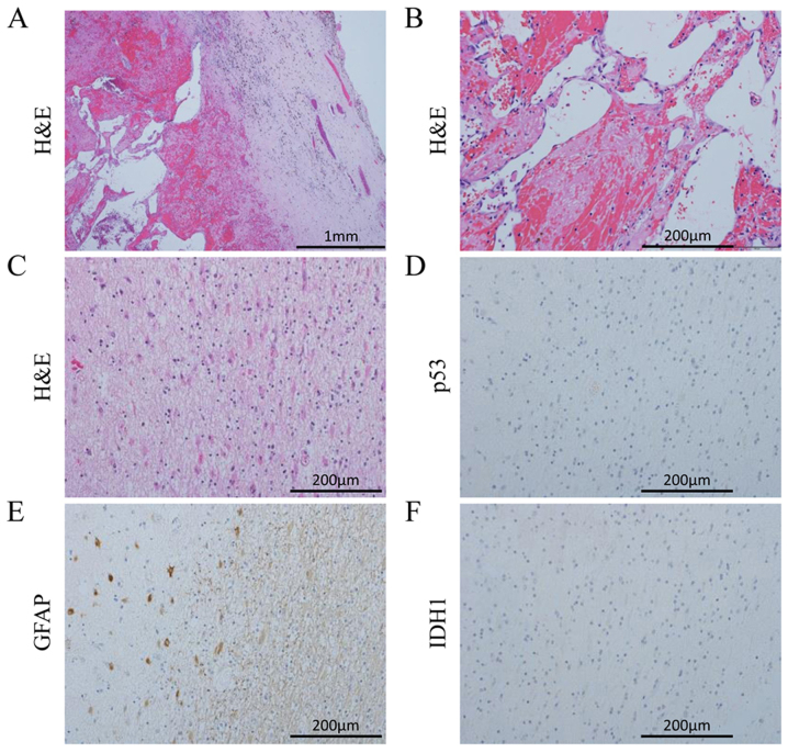
Figure 3.
Hemangioma sample obtained during the sixth surgery exhibiting (A) a connective tissue capsule and (B) sinusoidal vessels. In the autopsy specimens of the left frontal periventricular zone, GFAP-positive astrocytes were detected, but the cells were negative for p53 and IDH1 (C-F). H&E, hematoxylin and eosin; IDH1, isocitrate dehydrogenase 1; GFAP, glial fibrillary acidic protein.

